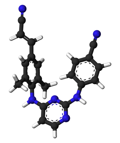
File:Rilpivirine 3D 2zd1.png
Appearance

Size of this preview: 505 × 600 pixels. Other resolutions: 202 × 240 pixels | 404 × 480 pixels | 647 × 768 pixels | 1,100 × 1,306 pixels.
Original file (1,100 × 1,306 pixels, file size: 197 KB, MIME type: image/png)
File history
Click on a date/time to view the file as it appeared at that time.
| Date/Time | Thumbnail | Dimensions | User | Comment | |
|---|---|---|---|---|---|
| current | 22:28, 20 May 2011 |  | 1,100 × 1,306 (197 KB) | Fvasconcellos | {{Information |Description= Ball-and-stick model of {{w|rilpivirine}} ('''TMC278''').<br />Created using [http://www.accelrys.com/products/downloads/ds_visualizer/index.html Accelrys DS Visualizer Pro 1.7] and {{w|GIMP}}. Optimized with {{w|OptiPNG}}. |
File usage
The following page uses this file:
Global file usage
The following other wikis use this file:
- Usage on fa.wikipedia.org
- Usage on hu.wikipedia.org
- Usage on it.wikipedia.org
- Usage on ko.wikipedia.org
- Usage on or.wikipedia.org
- Usage on ru.wikipedia.org
- Usage on sh.wikipedia.org
- Usage on sl.wikipedia.org
- Usage on sr.wikipedia.org
- Usage on uk.wikipedia.org
- Usage on vi.wikipedia.org
